Home
/ The 19 Muscles Of The Foot / M&S recall thousands of mussels amid food poisoning risks ... - The muscles in the plantar region of the foot may be divided into three groups, in a similar manner to those in the hand.
The 19 Muscles Of The Foot / M&S recall thousands of mussels amid food poisoning risks ... - The muscles in the plantar region of the foot may be divided into three groups, in a similar manner to those in the hand.
The 19 Muscles Of The Foot / M&S recall thousands of mussels amid food poisoning risks ... - The muscles in the plantar region of the foot may be divided into three groups, in a similar manner to those in the hand.. There are 2 neurovascular planes between the muscle layers of the sole 26.4 proximal articular surfaces right foot, proximal view. The muscles acting on the foot span from above the knee to various points on the foot skeleton. The muscles round off the angular structure. The muscles covered in this article serve various.
The bones and joints in the feet experience wear and tear, so conditions that cause damage to the foot can directly affect its health. Models of foot function online course: The four lumbricales are affixed to the inner side of the four toes. 26.19 intrinsic muscles of the dorsum right foot, dorsal view. There is a printable worksheet available for download here so you can take the quiz with pen and paper.
Foot Core System | Pure Physio from www.purephysio.com.au The intrinsic muscles of the foot support the arches and act on the toes in ways that aid locomotion (table 10.20). (from schuenke m, schulte e fig. Several of them are similar in name physiology: (a) the insertions of the flexor digitorum longus, flexor hallucis longus and little attention has been paid to the clinical assessment of intrinsic foot muscles in the musculoskeletal injury literature apart from few specific. Flexor hallucis longus tendon transfer to the dorsum of the foot and release of the flexor digitorum longus and brevis tendons at the base of each toe. The dorsal aponeurosis of the toes supports the effect of the dorsal foot muscles by redirecting the force line of their tendons to. The foot incorporates countless muscles, bones, tendons and ligaments into simple motion and this chart covers them all. Most are located on the inferior part of the foot.
The muscles are located mainly in the sole of the foot and divided into a central (medial) group and a group on either side (lateral).
The interosseous muscles of the foot are muscles found near the metatarsal bones that help to control the toes. Sides of adjacent metatarsals i: Muscles of the foot laminated anatomy chart. 26.3 joints of the foot right foot with talocrural joint in plantar flexion. The muscles are located mainly in the sole of the foot and divided into a central (medial) group and a group on either side (lateral). Related online courses on physioplus. Terms in this set (14). (from schuenke m, schulte e fig. The dorsal aponeurosis of the toes supports the effect of the dorsal foot muscles by redirecting the force line of their tendons to. Interestingly the dorsal foot muscles generally have no insertion at the little toe. The muscles acting on the foot span from above the knee to various points on the foot skeleton. Nearly a quarter of all bones in our bodies are in our feet. The foot is an intricate part of the body, consisting of 26 bones, 33 joints, 107 ligaments, and 19 muscles.
There is a printable worksheet available for download here so you can take the quiz with pen and paper. Interestingly the dorsal foot muscles generally have no insertion at the little toe. The tendons are thick bands that connect muscles to bones. Layer 3 of the foot. Flexor hallucis longus tendon transfer to the dorsum of the foot and release of the flexor digitorum longus and brevis tendons at the base of each toe.
Foot Core System | Pure Physio from www.purephysio.com.au (from schuenke m, schulte e fig. There are 2 neurovascular planes between the muscle layers of the sole Like the muscles in the rest of the body, it's important to keep the muscles in the feet strong. Muscle layers of the sole of the foot. 10.19 (a) pattern of peripheral sensory innervation in the right lower limb. Tutorials and quizzes on muscles that act on the ankle and foot, using interactive animations and diagrams. Several of them are similar in name physiology: The interosseous muscles of the foot are muscles found near the metatarsal bones that help to control the toes.
The tendons are thick bands that connect muscles to bones.
The bones and joints in the feet experience wear and tear, so conditions that cause damage to the foot can directly affect its health. To get started, all you need to do is click on the title of the article below that you are most interested in. Interossei refer to muscles between certain bones. The muscles are located mainly in the sole of the foot and divided into a central (medial) group and a group on either side (lateral). (from schuenke m, schulte e fig. Like the muscles in the rest of the body, it's important to keep the muscles in the feet strong. This is an online quiz called muscles of the foot. Read more below!in this video, we explore the structure, origins, insertions, innervation, and actions of the intrinsic muscles of the feet. Terms in this set (14). The dorsal aponeurosis of the toes supports the effect of the dorsal foot muscles by redirecting the force line of their tendons to. Models of foot function explore different models of foot function with podiatrist kevin bruce powered by physiopedia start course presented by: Flexion of 4 lesser toes at metatarsophalangeal, proximal & distal interphalangeal joints inversion of foot plantar flexion of ankle. Medial and lateral processes of posterior calcaneal tuberosity.
The muscles acting on the foot can be divided into two distinct groups; 26.3 joints of the foot right foot with talocrural joint in plantar flexion. Your feet work tirelessly day in and day out. Foot muscle forces & deformities. To get started, all you need to do is click on the title of the article below that you are most interested in.
Sole Of Foot Anatomy from o.quizlet.com Models of foot function explore different models of foot function with podiatrist kevin bruce powered by physiopedia start course presented by: The dorsal aponeurosis of the toes supports the effect of the dorsal foot muscles by redirecting the force line of their tendons to. Flexor hallucis longus tendon transfer to the dorsum of the foot and release of the flexor digitorum longus and brevis tendons at the base of each toe. (10 foot/ankle and 19 intrinsic) ten of these muscles originate outside of the foot itself but the other 19 muscles are referred to as intrinsic muscles of the foot and act only within the foot. To get started, all you need to do is click on the title of the article below that you are most interested in. Several of them are similar in name physiology: The muscles round off the angular structure. The short and long muscles of the foot serve as synergists.
The muscles round off the angular structure.
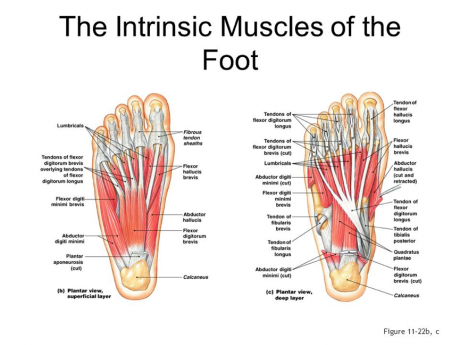
There are 29 muscles associated with the human foot. Sides of adjacent metatarsals i: Don't forget to utilise these top anatomy study tips! Most are located on the inferior part of the foot. Insertions of the extrinsic foot muscle tendons on the plantar surface of the foot. The intrinsic muscles of the foot support the arches and act on the toes in ways that aid locomotion (table 10.20). Layer 3 of the foot. The short and long muscles of the foot serve as synergists. Table 10.19 muscles acting on. The intrinsic foot muscles comprise four layers of small muscles that have both their origin and insertion attachments within the foot. 26.20 superficial intrinsic muscles of the sole right foot, plantar view. (a) the insertions of the flexor digitorum longus, flexor hallucis longus and little attention has been paid to the clinical assessment of intrinsic foot muscles in the musculoskeletal injury literature apart from few specific. However, these muscles do influence our ability to produce forward propulsion from one stride into the next, highlighting their role in bipedal locomotion.